National and Provincial Joint Engineering Research Centre for Marine Germplasm Resources Exploration and Utilization, Zhejiang Ocean University, Zhoushan 316022, China
2.
Marine Science and Technology College, Zhejiang Ocean University, Zhoushan 316022, China
Funds:
The National Natural Science Foundation of China under contract 42171069 and 41976121.
Despite most eel gobies (Gobionellidae: Amblyopinae) have inhabited brackish or marine waters, few species (such as Taenioides sp.) have been found to invade multiple inland freshwaters via artificial water transfer projects. The habitat transfers from brackish water to freshwater zones of Taenioides sp. have caused severe damage to Chinese aquatic ecosystems in recent years. Unfortunately, the molecular mechanism underlying freshwater invasion remains poorly understood. Considering changes of environment factors, especially salinity, are bound to adjust the demands for energy affected by mitochondria via oxidative phosphorylation, 13 Amblyopinae mitogenomes were compared, including the newly assembled Taenioides sp. mitogenome in this study. Comparative mitogenomic analyses revealed a highly conserved structure, composition and arrangements, with the exception of variable control region (CR). All of the CRs possessed tandem repeat sequences except Trypauchenopsis sp. G341, differing in motifs and number of copies, which was the dominant factor resulting in length heterogeneity of CR. The phylogenetic trees reconfirmed the paraphyletic origin of Amblyopinae with respect to Oxudercinae, supporting that these two subfamilies should be merged as an expansion of phenotypic variation within the “terrestrial goby” clade. Furthermore, four protein coding genes (COI, ND3, ND5 and Cyt b) in Taenioides sp. mitogenome have experienced adaptive evolution, indicating their important roles in enhancing the efficiency of ATP production to cope with the osmotic regulation adjustment and reach its current widespread distribution in multiple inland freshwaters of China. These results revealed the functional importance of mitochondrial genes, and provided fresh insights into the molecular mechanisms underlying the freshwater invasion. Also, our results may provide critical reference value for the future control of other invasive species.
Figure 1. Comparative analyses of 13 Amblyopinae mitogenomes. The intensity of the ring color indicates the degree of sequence conservation. The innermost ring to the outermost is presented as follows: Odontamblyopus rebecca, Odontamblyopus lacepedi, Odontamblyopus sp. ZL-2016, Trypauchenopsis sp. G341, Paratrypauchen microcephalus, Trypauchen vagina, Ctenotrypauchen chinensis, Amblyotrypauchen arctocephalus, Taenioides sp. CHY-2020, Taenioides gracilis, Taenioides anguillaris, Odontamblyopus rubicundus and Taenioides sp. XM-2018.
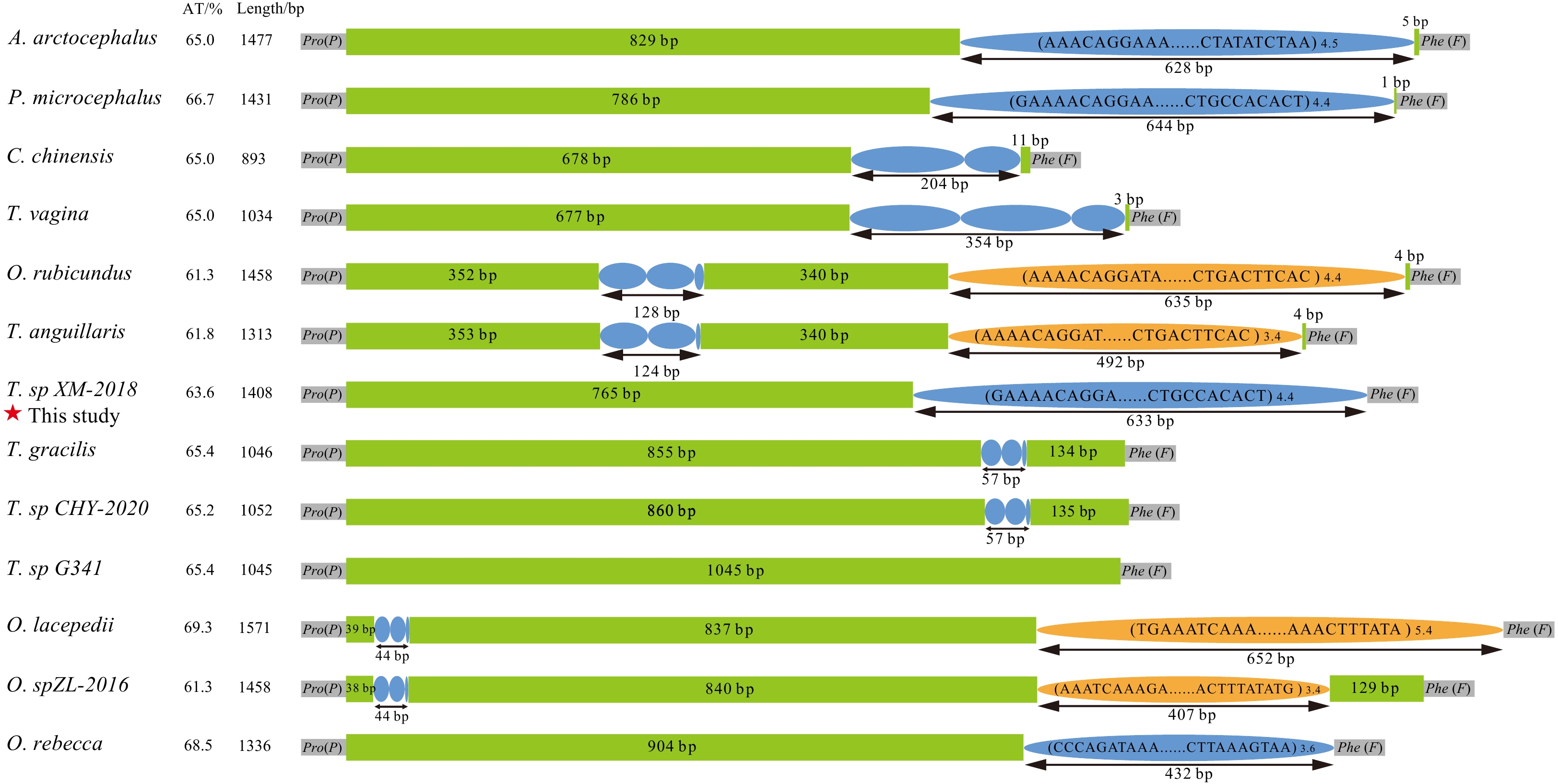
Figure 2. Comparative analyses of the organization of 13 Amblyopinae CRs. Purple and orange ellipses indicate the tandem repeat units; the remaining regions are shown in green boxes. The tandem repeat with copy number is displayed in the format of (motif) n.
Figure 3. Aligned sequences of 13 Amblyopinae CRs. The shaded blocks indicate the conserved sequences. Abbreviations of species names are given as follows: A.arc: Amblyotrypauchen arctocephalus; P.mic: Paratrypauchen microcephalus; C.chi: Ctenotrypauchen chinensis; T.vag: Trypauchen vagina; O.rub: Odontamblyopus rubicundus; T.ang: Taenioides anguillaris; T.sp1: Taenioides sp. XM 2018; T.gra: Taenioides gracilis; T.sp2: Taenioides sp. CHY 2020; T.sp3: Typauchenopsis sp. G341; O.lac: Odontamblyopus lacepedi; O.sp: Odontamblyopus sp. ZL-2016; O.reb: Odontamblyopus rebecca.
Figure 4. The phylogenetic relationship between Amblyopinae and their closely related Oxudercinae. The node labelled with a solid circle indicates 100 maximum likelihood bootstrap support and 100% Bayesian inference posterior probability. The numbers on the branches are bootstrap support for ML analyses (left) and posterior probability (right) for Bayesian inference.
Figure 5. The sequence alignment (a) and predicted three-dimensional protein structure of ND5 in freshwater-adapted Taenioides sp. and its closely related species inhabiting brackish and/or sea water (b). The positively selected sites were marked with red dotted box (a). Red squares represent the changes in the three-dimensional structure; numbers show the position of changes (b).


 DownLoad: